

PPCAR-NetResearch preview

PPCAR-Net: Projection-Refined Parametric
3D Coronary Artery Reconstruction
from Sparse X-ray Angiographic Views

From a few segmented projections to a branch-structured coronary artery — with smooth centrelines and detailed radius profiles.

Yu Ren1,2Hwee Kuan Lee1Tat-Jen Cham2Jonathan Yap3Khung Keong Yeo3

1 Bioinformatics Institute, Agency for Science, Technology and Research (A*STAR)2 College of Computing and Data Science, Nanyang Technological University3 National Heart Centre Singapore
SEGMENTED VIEWS
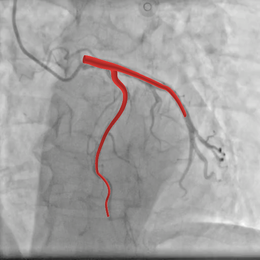
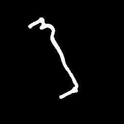
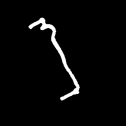
First segmented projection of RCA case 476Second segmented projection of RCA case 476

Two projections of the same artery

→

3D reconstruction

EXPLICIT 3D RECONSTRUCTION
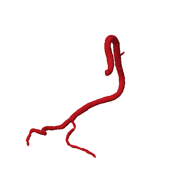
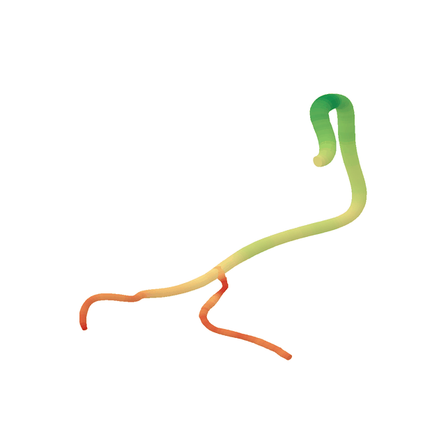
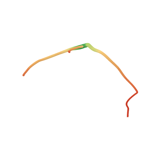
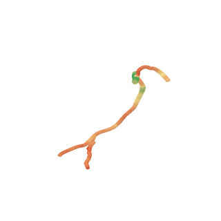
Rotating reconstruction of RCA case 476, colored by radius
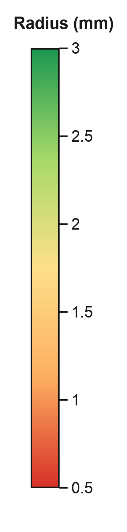
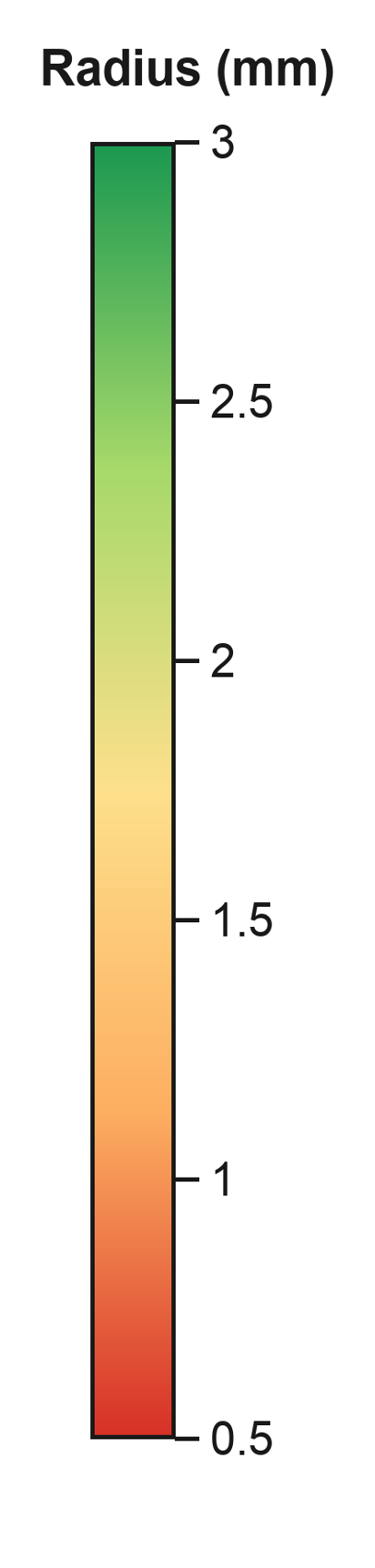
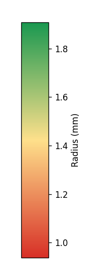
Radius (mm)Radius colour scale in millimetres for RCA case 476

CT-derived simulated projections and their predicted 3D anatomy. Colours in the reconstruction indicate local vessel radius.

ABSTRACT

Reconstructing 3D coronary anatomy from sparse views.

Sparse-view 3D coronary reconstruction commonly relies on cross-view correspondence and triangulation, which are vulnerable to vessel overlap and foreshortening, or on volumetric prediction followed by vascular-graph extraction, which does not directly provide centrelines and radii. We introduce PPCAR-Net, a projection-refined parametric coronary artery reconstruction network that directly predicts a branch-structured centreline-and-radius representation, termed vessel code, without explicit point matching, triangulation, or an intermediate volume.

From a variable number of segmented views, a coarse predictor combines frozen VGGT features with learned branch queries to estimate active branches, their B-spline centreline trajectories, and approximate dense radius profiles. Projection-guided geometry and radius refiners sample input view evidence and predict residual corrections learned with 3D supervision. We evaluate representation fidelity and sparse-view 3D reconstruction quantitatively and qualitatively. On simulated angiographic masks generated from CT-derived coronary anatomy, PPCAR-Net produces better connected artery reconstructions and achieves strong centreline accuracy, particularly for RCA, while maintaining competitive volumetric overlap. Coarse-to-fine inference takes 121 ms, enabling real-time reconstruction.

METHODS

How It Works

Sparse angiographic views can hide depth, overlap branches and shorten their apparent length. PPCAR-Net combines anatomical patterns learned across training cases with evidence from the input projections to recover a plausible 3D artery.

How we represent an artery

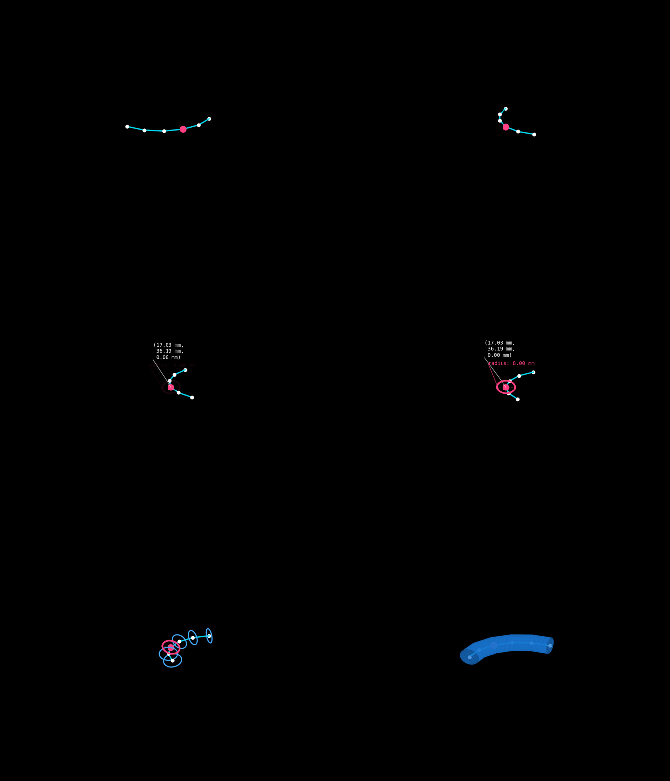
PPCAR-Net directly predicts vessel code, our explicit artery representation, which comprises branch presence, B-spline control points and dense radii. B-spline control points define each branch’s centreline, which we sample densely, with a radius associated with each sampled point. Learned refinement then adjusts the geometry and radius profiles using local projection evidence.

B-spline curvesContinuous centrelines within each branch
Animation illustrating B-spline centrelines and dense 3D sampling
Dense radiiLocal vessel thickness along the centreline
Animation showing how radius cross-sections along the centreline form the vessel surface

Predict the Structure,
Refine with the Evidence

PPCAR-Net uses a coarse-to-fine feed-forward pipeline to reconstruct an explicit 3D artery from calibrated coronary masks.

01

Predict the coarse vessel code

A frozen VGGT backbone extracts features from the input views. Learned branch queries combine these features with coronary anatomical patterns learned during training to predict a coarse vessel code: branch existence, the overall shape of each branch through B-spline centreline curves, and dense radius profiles.

02

Refine geometry and radii

We project the coarse prediction back into the input views and sample the corresponding local 2D evidence around the projected points. The refiners use this evidence to apply learned residual corrections to both the centreline geometry and the radii.

Two input views

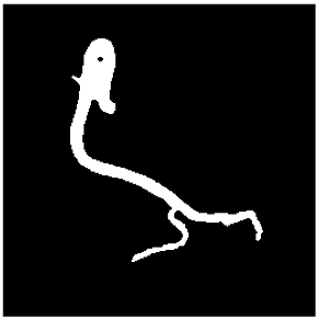
First segmented input view for the coarse-to-refined reconstruction exampleSecond segmented input view for the coarse-to-refined reconstruction example

Coarse prediction

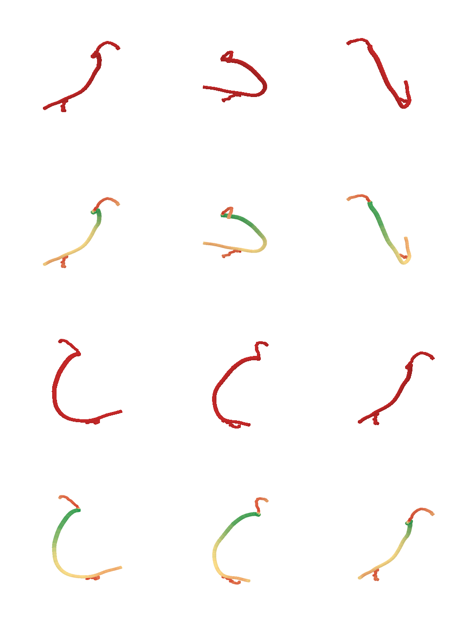
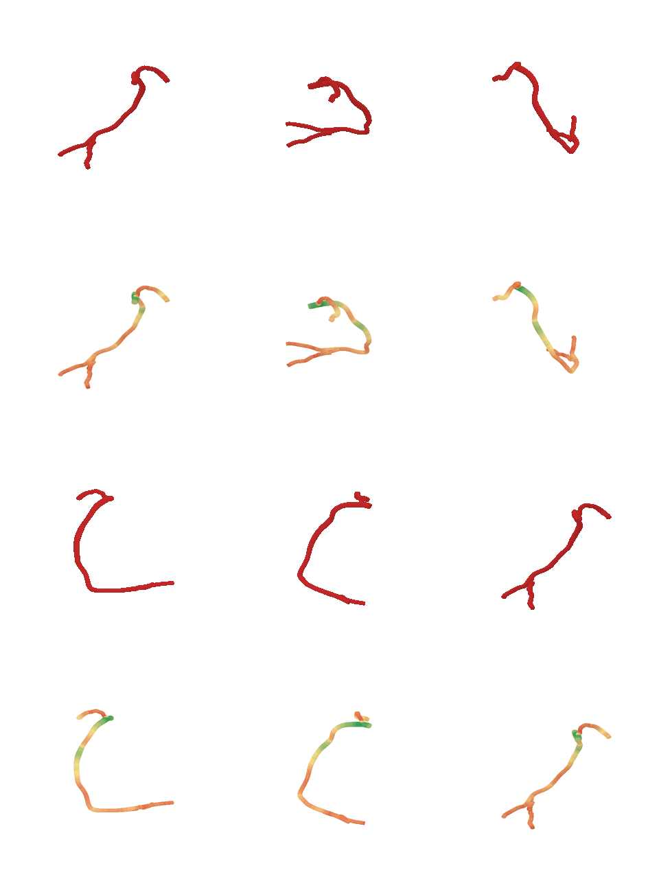
Coarse prediction: red vessel surface above and radius-coloured surface below

Refined prediction

Refined prediction: red vessel surface above and radius-coloured surface below
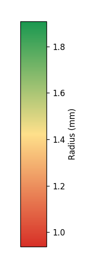
Shared radius colour scale in millimetres for the coarse and refined predictions
PPCAR-Net predicts a coarse vessel code from two segmented input views, then refines its geometry and radii. Each prediction shows the vessel surface in red (top) and coloured by local radius (bottom).
RESULTS

What It Reconstructs

Choose an artery and a case. Compare the CT-derived 3D label with the PPCAR-Net prediction from two segmented views.

Inference time

121 ms
Two-view coarse-to-fine inference · RTX 4090

Radius colours use the scale shown for each case. Rotations are pre-rendered visualisations; this page does not run model inference.

Browse the complete comparison gallery, including 1-, 2- and 4-view results ↗

The RCA and LCA examples use coronary CT angiography annotations from ImageCAS (Zeng et al.). The input masks are simulated projections of the annotated 3D arteries.

Diseased CT Anatomy

Recovering narrowing from 2D evidence

DISEASED CT ANATOMY · ASOCA

Two simulated projections of a diseased coronary artery, its 3D ground truth and our reconstruction.

Input projections

ASOCA segmented projection 1ASOCA segmented projection 2

3D ground truth

Rotating ASOCA ground-truth RCA mesh, mirrored to align with the prediction display

Our reconstruction

Rotating ASOCA prediction coloured by local radiusRadius colour scale from 0.5 to 3 millimetres

The ground-truth surface is shown in red; the prediction is coloured by local radius. The fixed views below highlight the local narrowing.

A closer look at the narrowing

Fixed views with the selected region circled and enlarged below.

Input projection 1

Magnified region

3D ground truth

Magnified region

Our reconstruction

Radius colour scale from 0.5 to 3 millimetres
Magnified region

A qualitative example of recovering visible radius reduction, rather than an evaluation of clinical stenosis detection.

This diseased coronary CT anatomy is from the ASOCA dataset (Gharleghi et al.), which provides CT coronary angiography images and artery annotations.

DOMAIN TRANSFER

On Real X-ray Angiography

Exploratory reconstruction from real angiographic images, using segmented artery masks as input.

From acquired views to 3D anatomy

REAL X-RAY EXAMPLE

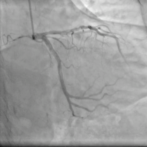
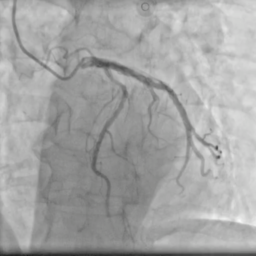
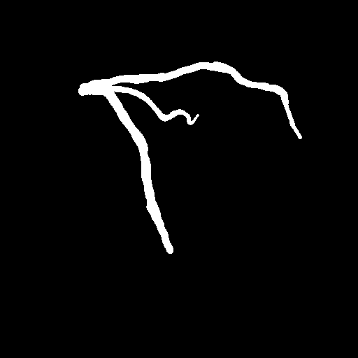
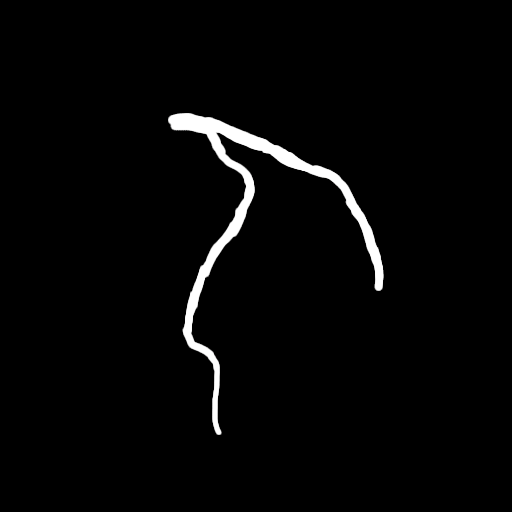
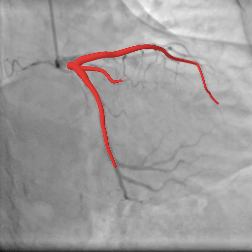
Acquired angiography and selected input frames are shown with their artery masks above; prediction overlays and the 3D reconstruction are shown below.

X-ray angiography

Selected views

Selected X-ray angiography frame, view 1Selected X-ray angiography frame, view 2

Artery masks

Segmented artery mask for real X-ray view 1Segmented artery mask for real X-ray view 2

First-view prediction overlay

Prediction overlay on real X-ray view 1

Second-view prediction overlay

Prediction overlay on real X-ray view 2

3D reconstruction

Rotating reconstruction from real X-ray angiography, coloured by radiusRadius colour scale from 0.5 to 3 millimetres

Radius-coloured prediction from the two views. Paired 3D ground truth is unavailable.

These real X-ray examples lack paired 3D ground truth for accuracy assessment and do not constitute systematic clinical validation.

The real X-ray angiography example is from the data released in the AutoCAR paper repository: Zhu et al., Sparse and transferable three-dimensional dynamic vascular reconstruction for instantaneous diagnosis (2025). Reconstructions shown here are produced by PPCAR-Net.

CITATION

BibTeX

If you use this work, please cite:

@misc{ren2026ppcarnet,
  title={{PPCAR-Net}: Projection-Refined Parametric 3D Coronary Artery Reconstruction from Sparse X-ray Angiographic Views},
  author={Ren, Yu and Lee, Hwee Kuan and Cham, Tat-Jen and Yap, Jonathan and Yeo, Khung Keong},
  year={2026},
  eprint={2610.09383},
  archivePrefix={arXiv},
  primaryClass={cs.CV},
  url={https://arxiv.org/abs/2610.09383}
}